Human cortical organoids cultured in vitro for 5 years; methylation-clock predicted age correlates with time in culture (r = 0.88–0.90)
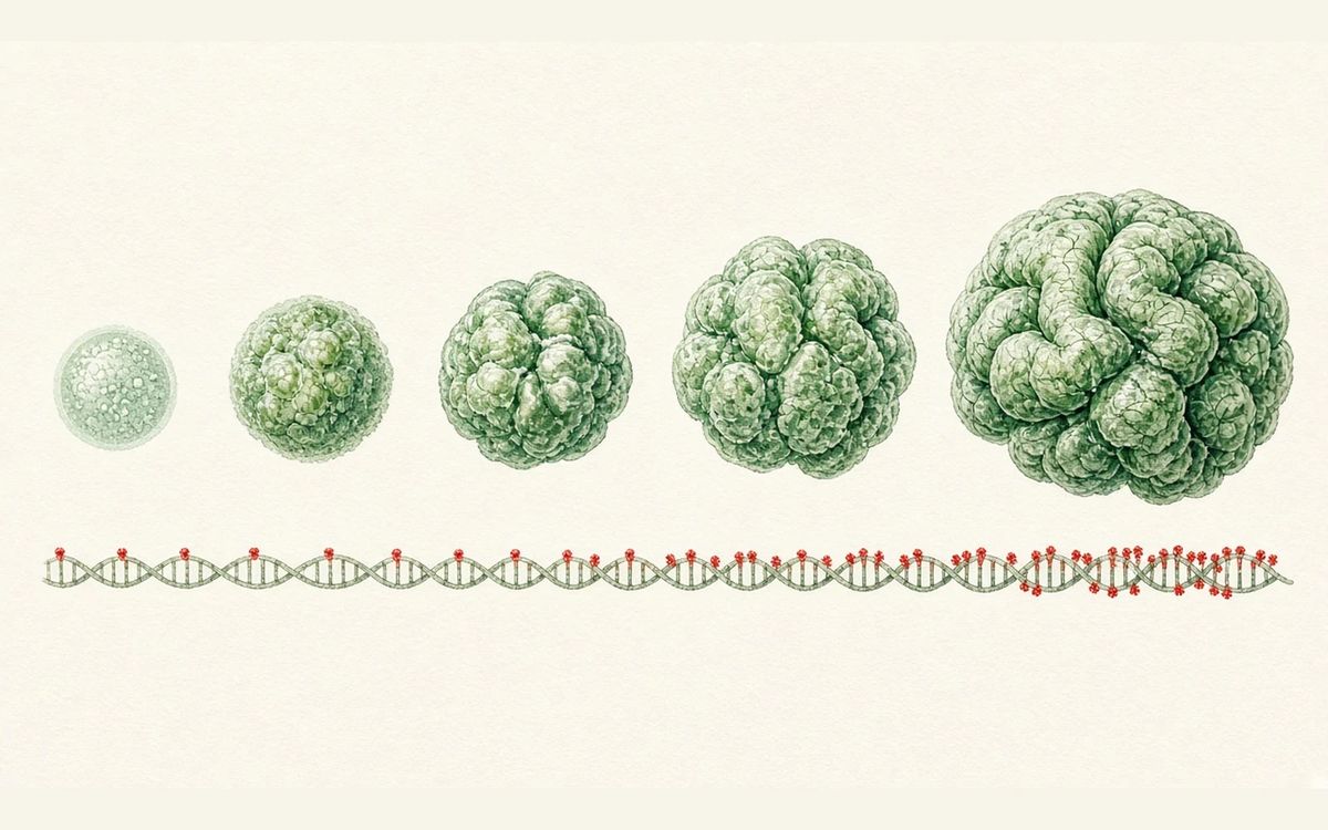
Human cortical organoids were cultured for up to 5 years, and ages predicted by the Horvath and cortical methylation clocks correlated with time in culture (r = 0.88–0.90, median absolute errors of 7.25 and 20.04 months).
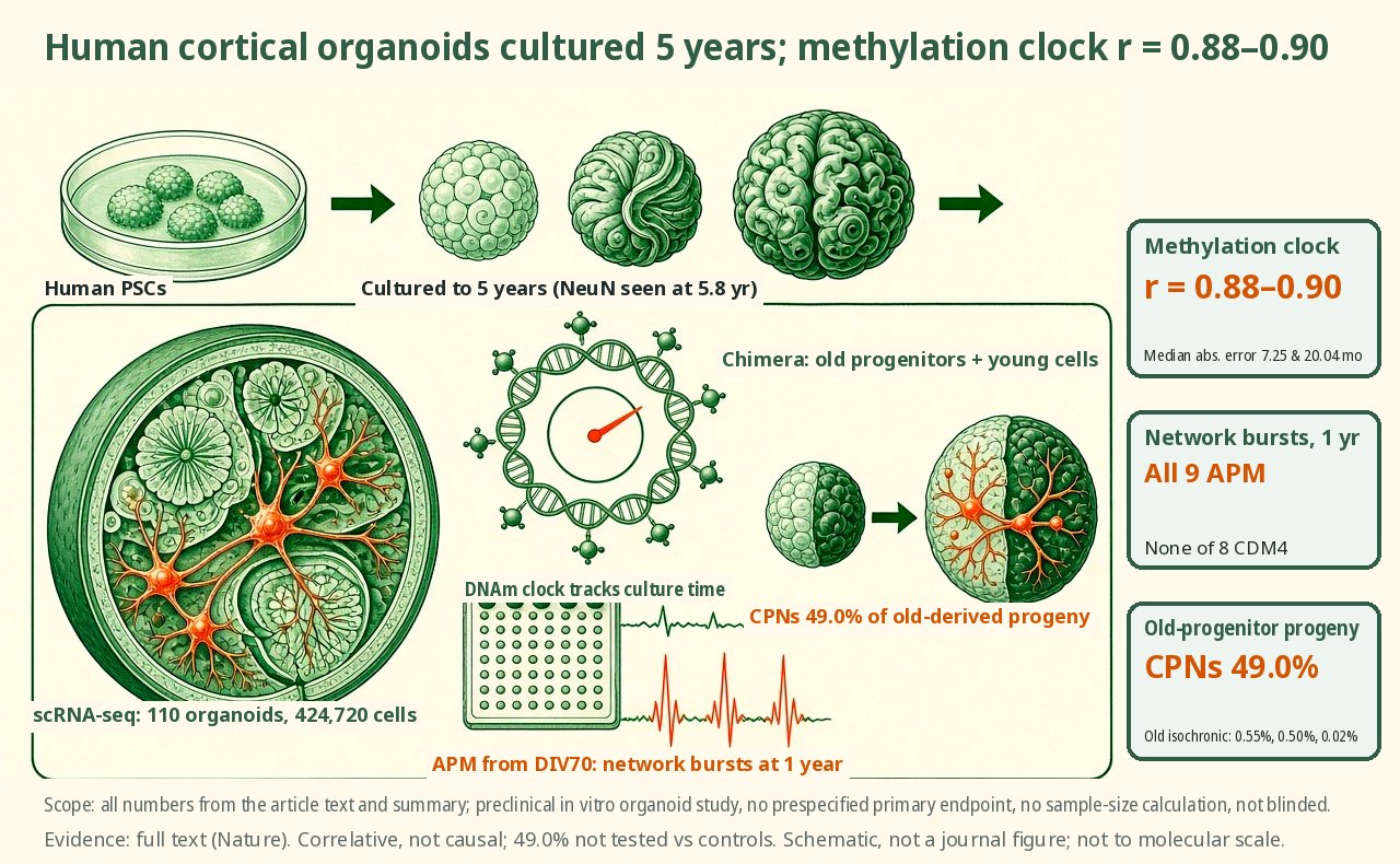
Human cortical organoids were cultured for up to 5 years (NeuN staining was seen at 5.8 years); scRNA-seq covered 110 organoids and 424,720 cells. Ages predicted by the Horvath and cortical methylation clocks correlated with time in culture (r=0.88–0.90), with median absolute errors of 7.25 and 20.04 months. After switching to APM from DIV70, SATB2+ cells increased at 9 months, and at 1 year none of 8 CDM4 organoids showed network bursts while all 9 APM organoids did. Transplanting old progenitors into young organoids produced late-born neurons directly, with CPNs making up 49.0% and early stages skipped, pointing to a cell-intrinsic developmental clock.
Key data card
- Study type: Preclinical in vitro study (human pluripotent stem cell-derived cortical organoids and chimeric organoids, Nature)
- Sample size n: scRNA-seq of 110 individual organoids (34 new, 76 previously published) and 424,720 cells; APM versus CDM4: 9 organoids each for SATB2 at 9 months, and 9 APM versus 8 CDM4 organoids for MEA at 1 year; 3,969 cells in heterochronic chimeras, three replicates for old isochronic chimeras, and n = 3 per group for SATB2 staining in the three groups
- Controls: Standard CDM4 medium; for the chimera experiments, old isochronic chimeras and 9-month standard organoids
- Intervention/dose: Switch to APM (BrainPhys plus GlutaMax) from DIV70; heterochronic chimeras: 9–12-month-old progenitors co-aggregated with DIV15 young cells
- Follow-up: Culture up to 5 years (NeuN immunostaining seen at 5.8 years)
- Primary endpoint: No protocol-specified primary endpoint; the core readout is the Pearson correlation and median absolute error between methylation-clock DNAm age and time in culture
- Primary endpoint result: Horvath pan-tissue clock and cortex-specific clock: r = 0.88–0.90, median absolute errors of 7.25 and 20.04 months; fetal brain clock r = 0.54, P = 0.056
- Statistics: LMM, NBME, Wilcoxon rank-sum, Fisher's exact test, ANOVA, one-sided F test, likelihood ratio test, Kruskal–Wallis; sample sizes were not calculated in advance and the study was not blinded
- Evidence level: Full text
- Verification record: Read the Europe PMC full text PMC13581598, including the Abstract, Main, Results subsections, Discussion, legends for Figs. 1–5 and Methods
- Cortical organoids cultured for 5 years, dated by transcriptome and methylation
- Switch to APM from DIV70, with network bursts still present at 1 year
- Old progenitors co-aggregated with DIV15 young cells
- CPNs made up 49.0% of progeny from old progenitors
Background and open questions
Human brain development and maturation span nearly two decades, and human-specific postnatal processes are hard to study in animal models. Human brain organoids provide an in vitro model, but most existing organoids recapitulate only early development; one earlier study cultured organoids to 694 days and analyzed them only by bulk RNA-seq and methylation arrays of whole organoids.
Long-term survival differs markedly between cell types, especially for neurons; beyond cell survival, it must also be shown that neuronal structure and circuit activity can be preserved over years in culture. Here the authors cultured cortical organoids for more than 5 years, integrating single-cell transcriptomics, whole-genome methylation, and structural and functional data, and used chimeric organoids to test whether progenitors retain a "memory" of the time they have experienced.
Study design
This is a preclinical organoid study with no prespecified primary endpoint, no power calculation or prespecified sample size for between-group comparisons, and no blinding. Cortical organoids were generated from several human pluripotent stem cell lines (such as H1, 11a and PGP1) following the authors' previous protocol. WGBS covered nine time points from 3 months to 5 years. Chimera experiments mixed 9–12-month-old progenitors with DIV15 young cells.
The scRNA-seq time course spanned 15 days to 5 years and included 110 individual organoids (34 new, 76 previously published) and 424,720 cells, mapped by label transfer onto human fetal and postnatal cortex reference datasets. From DIV70 onward, cultures were switched to activity-permissive medium (APM, BrainPhys plus GlutaMax), with standard CDM4 as the control; readouts included electron microscopy and 3D multielectrode arrays (MEA).
Key results
Transcriptomes match stages of human brain development
Transcriptionally, organoids at 15 days to 2 months corresponded to first-trimester fetal brain, 3 to 6 months mainly to the second trimester, and 9 months to 5 years shifted progressively toward late prenatal and postnatal stages, with postnatal-like features appearing after 12 months. Pearson correlations with time in culture for the DIALOGUE maturation modules MCP1–5 were −0.08, 0.78, 0.35, 0.78 and −0.72, respectively, with MCP4 used as the maturation score.
Methylation clocks correlate with time in culture
Epigenetically, WGBS covered nine time points from 3 months to 5 years. DNAm ages predicted by the Horvath pan-tissue clock and the cortex-specific clock correlated closely with time in culture (r = 0.88–0.90), with median absolute errors of 7.25 and 20.04 months; the fetal brain clock showed only a positive, non-significant correlation (r = 0.54, P = 0.056). Methylation at solo-WCGW sites remained stable, indicating that these changes are not driven mainly by cell proliferation.
APM preserves neurons and synapses
Under standard CDM4 the proportion of neurons declined with time in culture, and the 5-year sample retained only a small mixed neuronal cluster of 101 cells. After switching to APM from DIV70, SATB2+ cells increased at 9 months (9 organoids per condition, LMM P = 2.4×10⁻⁵). Electron microscopy showed no difference in synapse density at 6 months (P = 0.7) but higher density with APM at 1 year (P = 0.002); the proportion of synapses on spines was 25% (CDM4) versus 52% (APM) (Fisher P = 9.5×10⁻⁸).
Maturation scores and network activity
Maturation scores for excitatory neurons were higher overall with APM (one-sided F test, P = 4.03×10⁻²); by time point, P = 1.56×10⁻³ at 4 months, P = 9.77×10⁻¹ at 6 months and P = 3.86×10⁻⁶ at 1 year. 3D MEA recordings showed that at 1 year none of 8 CDM4 organoids displayed network bursts while all 9 APM organoids did; 2-year APM organoids still burst, and at least two neuronal subpopulations could be distinguished.
Old progenitors skip early fates
In human heterochronic chimeric organoids, CPNs made up 49.0% of the progeny of 9–12-month-old progenitors, versus 0.55%, 0.50% and 0.02% in three old isochronic chimera replicates (the third at 11 months) and 1.1% in 9-month standard organoids; they appeared within about 2 weeks of re-aggregation. SATB2 immunostaining across the three groups (n = 3 each) gave Kruskal–Wallis P = 0.0519, not significant.
Mechanistic interpretation
Demonstrated in the paper: The correlations of transcriptional maturation score and methylation-clock age with time in culture were measured directly and are correlative evidence. APM cultures had more SATB2+ cells at 9 months, higher synapse density and a higher proportion of spine synapses at 1 year, and retained network bursts; whether activity is a causal mediator was not tested here by intervention.
The heterochronic chimera experiment directly shows that after co-aggregation with young cells, old progenitors had lower maturation scores than progenitors kept in old isochronic culture, and that their progeny were 49.0% CPNs, while young-derived cells in the same chimera produced only early cell types. This suggests old progenitors can respond to inductive signals from young cells.
Author hypotheses: The authors propose that old progenitors retain a "memory" of the developmental steps they have already completed, so that on exposure to early inductive signals they still skip early progeny and directly generate excitatory neurons that normally require months of culture. They also suggest that promoting spontaneous activity benefits neuronal survival and maturation, and that adding in vivo factors such as sensory-evoked activity and non-neural tissue could improve organoids further.
Limitations and uncertainties
- Under CDM4 the 5-year sample retained only a small mixed neuronal cluster of 101 cells, too few to subdivide; staining specificity declined after 1 year, limiting antibody interpretation, and 2- to 3-year samples required a switch to a fresh-frozen protocol; heterochronic chimeras succeeded at very low rates.
- At 1.5 years each group had only n = 2, and 60-month scRNA-seq had only n = 2, with no prespecified sample sizes; in the MEA experiments the CDM4 organoids were switched to APM only 2 weeks before recording, so the two groups did not share identical culture histories.
- The 9-month maturation score difference was P = 5.88×10⁻², yet the main text states that all time points except 6 months were significant; the three-group SATB2 comparison gave P = 0.0519; the fetal brain clock gave P = 0.056; the cortical clock had a median absolute error of 20.04 months; and the 49.0% figure was not formally tested against controls.
- All results come from in vitro organoids with no direct functional comparison to postnatal human brain tissue, so extrapolation to in vivo maturation warrants caution.
Clinical and industry implications
If these results can be reproduced in more donors and larger samples, long-term organoid cultures could become an in vitro platform for studying human postnatal cortical maturation and for testing timekeeping mechanisms. APM as a culture condition may serve as a reference for long-term experiments that need excitatory neurons preserved.
The progenitor time-memory results suggest that heterochronic chimera designs can be used to probe fate potential in organoids, though this remains an in vitro observation. The authors also note that further integration of sensory-evoked activity and non-neural tissue is a direction for improving organoids. No data on patients or treatment endpoints are involved at this stage.
Authors, source and verification
Evidence level: Full text; verification record: Read the Europe PMC full text PMC13581598, including the Abstract, Main, Results subsections, Discussion, legends for Figs. 1–5 and Methods
Faravelli I, Antón-Bolaños N, Wei A, Faits T, Kumar AS, Andreadis S, et al. Human brain organoids record the passage of time over multiple years. Nature. 2026. https://doi.org/10.1038/s41586-026-10877-x
Primary field: Organoids · Related: Long-term culture, DNA methylation clocks, Single-cell transcriptomics, Chimeric organoids, Activity-permissive medium, 3D multielectrode arrays
Summary of a published paper or preprint, written from the original text; numbers are as reported by the authors. Not medical or investment advice. Corrections: contact@
Related science
TSC2 loss produces astrocyte reactivity in organoids at all 3 time points
Astrocytes in a brain organoid cross-section, with reactive cells in red
Human cortical organoids in apallial mice: grafts make up 91.9% of cortical volume
The green area in a brain cross-section is the transplanted human cortical organoid
Transient confinement culture gives small intestinal organoids a 100% engraftment rate with early transplantation
Ring-shaped small intestinal organoids in a dish, with one on the right engrafted into the intestinal wall
One email, with links to every paper. Reports and custom landscapes: contact@inlightbio.com.