Human cortical organoids transplanted into apallial mice: grafts make up 91.9% of cortical volume and grow about 4.7-fold between 2 and 3 months
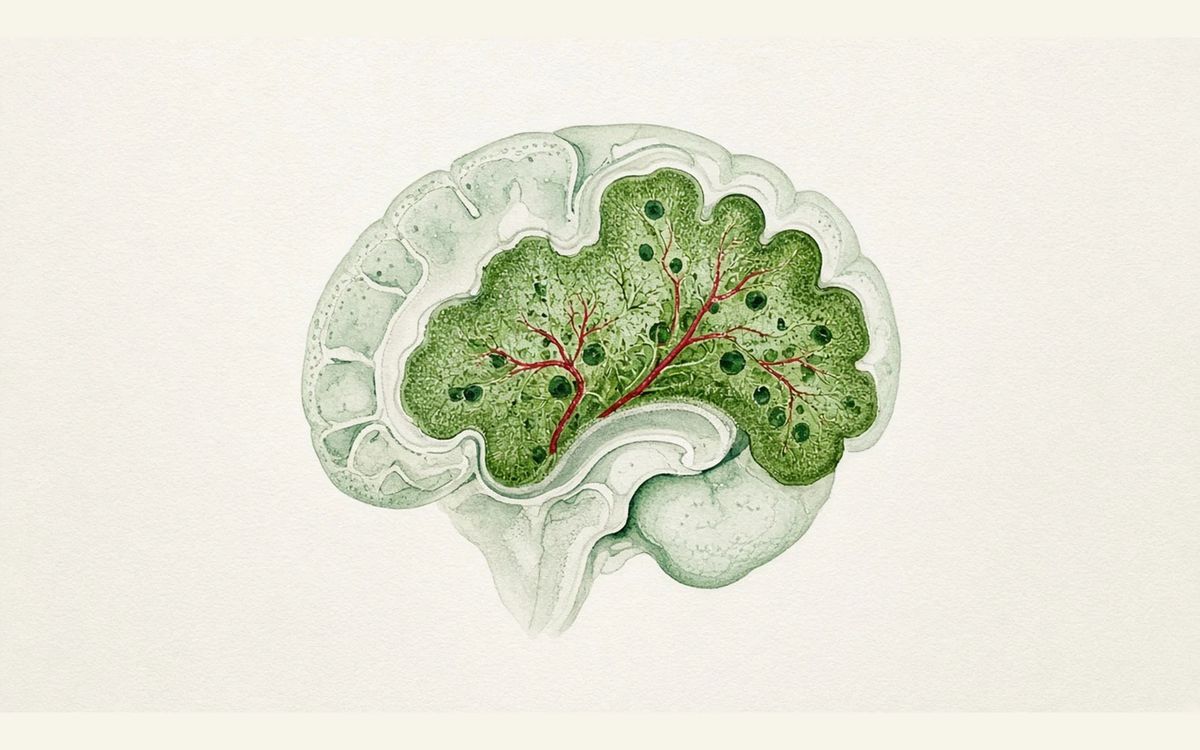
Four human cortical organoids were transplanted neonatally into immunodeficient apallial mice lacking Esco2, and by 3 months the grafts made up 91.9% of cortical tissue volume, growing about 4.7-fold between 2 and 3 months.
In immunodeficient apallial mice lacking Esco2, 2 human cortical organoids were transplanted into each hemisphere (4 per animal) at 5–17 days of age. The transplantation success rate was 86.2% (29 mice), grafts grew about 4.7-fold between 2 and 3 months (n=14, paired t test P=1.66×10⁻⁶), and at 3 months they made up 91.9% of cortical tissue volume (n=7). Calcium imaging showed graft-wide synchronous calcium bursts lasting tens of seconds and recurring every few minutes. Grafts contained L5-ET neurons, and sparse human projections were seen in the cervical spinal cord. In the Y maze, transplanted mice showed spontaneous alternation above chance (P=0.0004) while apallial mice did not.
Key data card
- Study type: Preclinical animal study (human iPS cell-derived cortical organoids xenotransplanted into mice)
- Sample size n: 29 mice for the transplantation success rate; n = 14 for growth; n = 7 for cortical volume fraction; 2 mice per group for whole-brain snRNA-seq; 3 mice each for calcium imaging and LFP; CatWalk with 16 controls, 10 apallial and 7 XCX; Y maze with 39 controls, 16 apallial and 18 XCX; hypoxia CatWalk with 6 female and 5 male controls, 9 female apallial, and 4 female and 5 male XCX
- Controls: Littermate Esco2fl/fl Prkdcscid/scid control mice; focal grafts transplanted into intact cortex; wild-type mice; untransplanted apallial mice
- Intervention/dose: 2 hCOs transplanted into each hemisphere of apallial mice at 5 to 17 days of age, 4 per animal; the hypoxia group was exposed to 5% O2 for 5 hours (including a 1-hour ramp-down)
- Follow-up: MRI monitoring 2 to 3 months after transplantation; up to 177 days after organoid differentiation
- Primary endpoint: Graft volume growth measured by MRI (2 to 3 months after transplantation) and the fraction of cortical tissue volume occupied
- Primary endpoint result: About 4.7-fold growth between 2 and 3 months (n = 14, paired t test, t = 8.219, P = 1.66×10⁻⁶); 91.9% of total cortical tissue volume at 3 months (n = 7, descriptive); transplantation success rate 86.2% (29 mice)
- Statistics: Paired/unpaired t tests, ANOVA (Holm–Šidák correction), Kruskal–Wallis (Bonferroni correction) and one-sample t tests, all two-sided
- Safety: Apallial mice weighed less than controls and showed a trend toward lower survival at about 2 months of age; 1 mouse died of airway obstruction during hypoxia induction
- Evidence level: Full text
- Verification record: Read the Nature open-access full-text HTML abstract, Results, Discussion, legends for Figs. 1–5 and Methods
- Emx1-cre deletion of Esco2 eliminates dorsal cortex
- Four hCOs transplanted per animal neonatally
- MRI tracks graft growth and volume fraction
- Detection of L5-ET neurons and spinal projections
- CatWalk gait readouts after hypoxia
Background and open questions
Human brain tissue is hard to obtain directly, and human stem cell-derived cortical organoids (hCOs) can model cortical neurogenesis but lack circuit- and behaviour-level readouts in vitro. Transplanting hCOs into neonatal rat cortex has previously promoted integration and maturation of human neurons, but grafts occupied at most about one third of one rat cortical hemisphere.
The paper argues that mouse neurons develop faster than human neurons, and that a rapidly maturing host environment and pre-existing rat connectivity may limit graft proliferation and projection, which matters especially for modelling neurodevelopmental disorders involving widespread circuit dysregulation and behavioural change. This study's approach is to genetically eliminate dorsal and medial cortex in mice first, then transplant hCOs into the vacated cortical cavity.
Study design
Apallial mice were generated on an immunodeficient SCID background by deleting Esco2 in Emx1-expressing cells (Emx1-cre+/− Esco2fl/fl Prkdcscid/scid), killing dividing cells. Apallial offspring made up 23.4% (n = 91 litters, 46 breeding cages), consistent with the 25% Mendelian expectation; MRI showed about 50% less whole-brain tissue than Esco2fl/fl Prkdcscid/scid controls.
Apallial mice aged 5 to 17 days (median 10 days) received 2 hCOs per hemisphere, 4 per animal, derived from 3 hiPS cell lines, and are termed xenocorticated (XCX) mice after transplantation. Controls were littermate Esco2fl/fl Prkdcscid/scid mice and focal grafts transplanted into intact cortex. The paper states that sample sizes were estimated empirically from previous studies and that the study was not powered to detect small sex effects.
Key results
Primary readout: graft growth and volume fraction
The transplantation success rate was 86.2% (29 mice, 3 hiPS cell lines), consistent with the earlier rat t-hCO protocol and indicating that hCO survival does not require an intact host cortex. MRI showed grafts growing about 4.7-fold between 2 and 3 months after transplantation (n = 14, paired t test, t = 8.219, P = 1.66×10⁻⁶). At 3 months, grafts made up 91.9% of total cortical tissue volume (n = 7), averaging about 32,000 neurons/mm3; this fraction is a descriptive value.
Extent of cortical ablation and L5-ET neurons
Whole-brain snRNA-seq (880,149 nuclei, 2 mice per group) showed roughly sevenfold reductions across classes of dorsal cortex-derived glutamatergic neurons, with olfactory bulb, non-cortical, amygdalar and piriform cortex-derived classes preserved, so "apallial" is only an operational label. In XCX mice, 85% of layer 5 extratelencephalic (L5-ET) neurons had UCell scores >0.1, versus only 3 of 213,753 neurons in the HNOCA organoid atlas; the L5-ET proportion in XCX was more than threefold higher than in previous rat and adult mouse transplants.
Behaviour: MoSeq and CatWalk
MoSeq (60 syllables, 99% of variance) placed XCX mice between controls and apallial mice but closer to apallial, with the greatest session-to-session drift (Kruskal–Wallis P = 0.019; XCX versus control Bonferroni-corrected P = 0.027), so transplantation did not normalize them. CatWalk showed no difference in run speed (ANOVA P = 0.8467); swing time was shorter in apallial mice than controls (P = 0.0019), with XCX versus control at P = 0.2303; diagonal paw synchrony was altered in both apallial and XCX groups.
Y maze and fear conditioning
In Y maze spontaneous alternation, controls (n = 39, one-sample t test against the 50% chance level P = 2.08×10⁻⁷) and XCX mice (n = 18, P = 0.0004) performed above chance while apallial mice (n = 16, P = 0.3016) did not; the effect replicated in two independent cohorts months apart. Apallial and XCX mice both showed reduced freezing during trace fear conditioning training and cued testing, along with contextual freezing deficits.
Cellular and gait readouts after hypoxic injury
After XCX and control mice were exposed to 5% O2 for 5 hours (including a 1-hour ramp-down), HIF1α immunoreactivity was visible in the human grafts of 3/3 XCX mice, with no clear signal in adjacent host archicortex or in identically exposed control mice. In CatWalk testing, XCX mice showed the largest increase from baseline in the proportion of 3- to 4-paw support (ANOVA P = 0.0047; control versus XCX P = 0.0120, XCX versus apallial P = 0.0076), while mean speed did not change (P = 0.3845).
Mechanistic interpretation
Demonstrated in the paper: XCX grafts at 4 months after differentiation (labelled with hSYN1-GCaMP8s) showed graft-wide synchronous calcium bursts lasting tens of seconds and recurring every few minutes, propagating across the entire dorsal surface within about 100 ms; bursts correlated with orofacial movement (paired t test P = 0.021, n = 3 mice), and 32-channel probe recordings captured LFP bursts occurring synchronously along the full extent (n = 3 mice).
All 3 XCX mice contained VEN-like bipolar or spiral cells, making up 0.16% of labelled somata, with a median soma size of 29.8 µm versus an overall median of 9.251 µm; sparse GCaMP8s+STEM121+ human projections were seen in the cervical spinal cord in all 3 XCX mice, with no signal in the spinal cords of focal graft controls or wild-type controls.
Author hypotheses: The authors suggest that genetic ablation of host cortex may allow enrichment of L5-ET neurons, including corticospinal neurons; behavioural output may reflect interaction between human grafts and preserved mouse subcortical circuits, but whether grafts are necessary or sufficient for specific behaviours remains unclear. Gait changes after hypoxia may be driven by longer hindpaw contact times and larger contact areas, an interpretation that was not tested causally.
Limitations and uncertainties
- Grafts reached at most 177 days after differentiation, with a transcriptional age at 24 weeks roughly equivalent to the late second trimester, lacking canonical cortical lamination, full regionalization, sufficient GABAergic interneurons and a mature transcriptional profile; the authors suggest that a mismatch in maturation rate between host and graft may limit integration.
- The study included no non-cortical organoid transplantation control, so cortical lineage effects cannot be distinguished from general transplantation effects; whether grafts are necessary or sufficient for specific behaviours remains unclear, and functional integration requires pathway-specific recording and manipulation.
- Whole-brain snRNA-seq used only 2 mice per group, and calcium imaging and LFP 3 each; VEN-like cells made up only 0.16% and were identified morphologically; the apallial group in the hypoxia behaviour experiment was female only (n = 9); the 91.9% fraction (n = 7) and the fold enrichment of L5-ET neurons were not statistically tested, and MoSeq also discriminated apallial from XCX mice only weakly.
Clinical and industry implications
If these results are reproduced across more cell lines and at later maturation time points, xenocorticated mice could provide a platform for assessing human cortical neurons in vivo in which grafts make up 91.9% of cortical tissue volume, while reading out circuit activity and behaviour at the same time. The gait changes in the hypoxia model suggest the platform could be used to model perinatal hypoxia and related neurodevelopmental disorders and to test therapeutic interventions on larger volumes of human cortical tissue.
The authors also note that if grafts become more mature, ethical guidance should be established early. All of the above are preclinical mouse data with no patient-level validation.
Authors, source and verification
Evidence level: Full text; verification record: Read the Nature open-access full-text HTML abstract, Results, Discussion, legends for Figs. 1–5 and Methods
Kaganovsky K, Kelley KW, Gschwind T, Harary PM, Kochalka J, White AD, et al. Developmental xenocortication using human-derived organoids in mice. Nature. 2026. https://doi.org/10.1038/s41586-026-11032-2
Primary field: Organoids · Related: Disease models, Xenotransplantation, Apallial mice, Cortical organoid transplantation, L5-ET neurons, MoSeq behavioural analysis
Summary of a published paper or preprint, written from the original text; numbers are as reported by the authors. Not medical or investment advice. Corrections: contact@
Related science
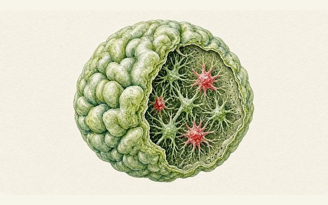
TSC2 loss produces astrocyte reactivity in organoids at all 3 time points
Astrocytes in a brain organoid cross-section, with reactive cells in red
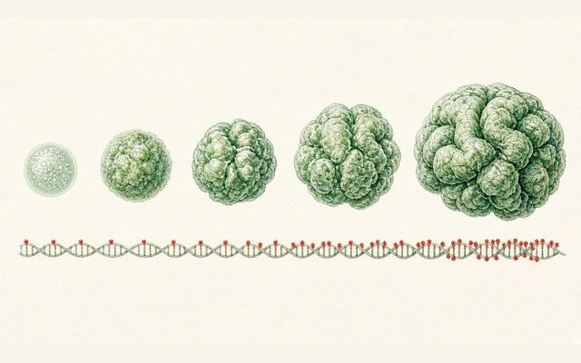
Cortical organoids at 5 years: methylation clocks track time with r=0.88–0.90
Organoids growing progressively larger, with red dots on the DNA strand below marking methylation sites
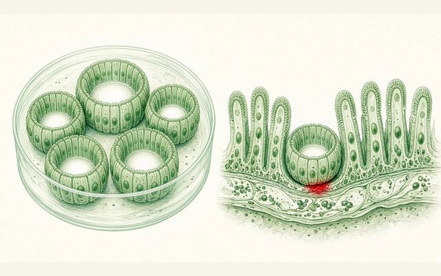
Transient confinement culture gives small intestinal organoids a 100% engraftment rate with early transplantation
Ring-shaped small intestinal organoids in a dish, with one on the right engrafted into the intestinal wall
One email, with links to every paper. Reports and custom landscapes: contact@inlightbio.com.