Transient confinement culture gives small intestinal organoids a 100% engraftment rate with early transplantation
In human small intestinal organoids, CCS transiently fused about 4,000 spheroids, achieving 100% engraftment on transplantation at d14, significantly higher than conventional d14 HIOs.
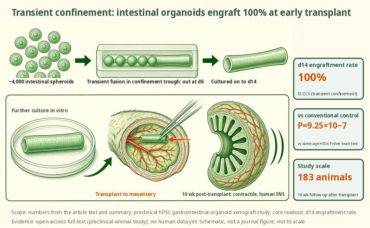
A confinement culture system transiently fused about 4,000 intestinal spheroids, which were removed on day 6, cultured on to day 14 and then transplanted onto the mesentery, giving a 100% engraftment rate, higher than conventional human intestinal organoids of the same age (exact test P of 9.25×10⁻⁷). The study used 183 animals in total, with 10 weeks of follow-up after transplantation, producing contractile intestinal tissue containing a human enteric nervous system. Conventional small intestinal organoids of the same age engrafted at a significantly lower rate.
Key data card
- Study type: Open-access full-text preclinical study of human hPSC-derived gastrointestinal organoids and xenotransplantation
- Sample size n: Size measurements in HIO n=6 and SI CCS n=9; unstimulated organ bath in tHIO+ENS n=4, SI CCS n=5 and adult small intestine n=3; 183 animals across the study
- Controls: Conventional HIO/HCO/HaGO, HIO+ENS, sham, and adult small intestine samples
- Intervention/dose: Transient fusion in PDMS confinement troughs; about 4,000 intestinal spheroids removed at d6 and cultured on to d14 before transplantation
- Follow-up: 10 weeks after transplantation; 12 weeks after tie-in
- Primary endpoint: Preclinical core readout: engraftment rate of SI CCS transplanted at d14, compared with conventional d14 HIOs transplanted onto the mesentery
- Primary endpoint result: SI CCS achieved 100% engraftment when transplanted at d14; versus d14 mesentery HIOs, Fisher's exact test P=9.25×10⁻⁷
- Statistics: Fisher's exact test, Mann–Whitney, ANOVA/Tukey and t tests for the key comparisons shown in the figures
- Evidence level: Full text
- Verification record: The source was the Nature Biomedical Engineering open-access full-text HTML material provided by the user; verification covered the Abstract, Results, Methods, figure legends and Discussion
- About 4,000 spheroids loaded into confinement troughs
- Fusion into a band from d0 to d5
- Removal at d6 and culture to d14
- Transplantation yields intestinal tissue with an ENS
Background and open questions
Human gastrointestinal organoids can model epithelium, mesenchyme and some functions, but conventional HIOs usually grow as spherical units far removed in shape from the intestinal tube; insufficient maturation before transplantation and limited size also constrain subsequent surgery and functional readouts. Conventional culture of colonic and gastric organoids also frequently suffers from low engraftment rates and small size.
Another bottleneck is the ENS. Existing assembloid strategies require neural crest cells and intestinal spheroids to be prepared separately and then recombined, making proportion and timing harder to control. Poling et al. take a physical-confinement approach: rather than adding neural cells separately, they let large numbers of spheroids co-develop transiently in long troughs to promote maturation.
Study design
This is an open-access full-text preclinical engineered-organoid study. The authors made PDMS confinement troughs from printed moulds. About 4,000 spontaneous spheroids were loaded into the troughs, still dispersed at d0 and fused into a unified structure by d5, then removed at d6 and cultured on in Matrigel to d14. SI CCS were then transplanted and compared with conventional HIOs, HIO+ENS and adult small intestine samples.
The animal design used 1 mouse per conventional organoid transplant and 1 rat per CCS transplant, with 183 animals used across the study. This is not a clinical trial and no between-group superiority power calculation is stated; key comparisons rely on Fisher's exact, Mann–Whitney, ANOVA or t tests. Neuromuscular readouts were quantified as the AUC over 60 s before and after stimulation, and cholinergic and nitrergic contributions were inferred from differences after sequential treatment with L-NAME and atropine.
Key results
Earlier transplantation
Unlike previous HIOs, which required at least 28 d of culture before kidney capsule or mesenteric transplantation, SI CCS could be transplanted at d14 with a 100% engraftment rate; versus d14 mesenteric HIOs, Fisher's exact test gave P=9.25×10⁻⁷. This approach replaces spherical units with long confined cultures, and on that basis the authors position SI CCS as tissue that can enter transplantation earlier.
Larger scale
The abstract states that CCS products can be up to 10 times larger than those from conventional methods; small intestinal CCS grafts reached 8 cm in width after 10 weeks, alongside higher engraftment rates. Size analysis included HIO n=6 and SI CCS n=9, and staining for proliferation, brush border enzymes and secretory cells supported structural maturation, with large stretches of continuous epithelium visible.
Ruling out host body size
To rule out host body size, the authors transplanted d28 HIOs into both mice and rats and saw no significant size difference at collection. Conventional organoids were thereafter placed mainly in mice and CCS mainly in rats, on that basis separating host size from graft-intrinsic effects.
Neuromuscular function
In the unstimulated state, organ baths compared tHIO + ENS (n=4), SI CCS (n=5) and adult small intestine (n=3); SI CCS did not differ significantly from adult small intestine. EFS evoked contraction in SI CCS, and TTX significantly reduced the response; bethanechol produced dose-dependent contraction and scopolamine induced relaxation, with all data normalized to tissue mass.
Extension and luminal exposure
Before transplantation, early progenitor subpopulations made up about 40% of neural clusters in d28 HIO + ENS and more than 80% in d14 SI CCS. CCS also extended to colon and stomach: after 10 weeks, C CCS reached about 6 cm in width, and G CCS showed improved engraftment and size. In an SI CCS "tie in" versus sham comparison, TEER did not differ significantly but paracellular permeability was significantly reduced; colonic and gastric CCS also showed ENS-dependent contraction in organ baths, and regional markers supported their tissue-type identity.
Mechanistic interpretation
Demonstrated in the paper: Immunostaining of 10-week SI CCS showed UCHL1-, TUBB3- and HuC/HuD-positive nerve bundles between the muscle layers, co-localized with XRCC5, supporting a human graft origin rather than host ingrowth; CHAT and NOS1 identified excitatory cholinergic and inhibitory nitrergic neuronal subtypes.
In organ baths, EFS evoked contraction in SI CCS and TTX significantly reduced the AUC; sequential inhibition with L-NAME and atropine showed that both nitrergic and cholinergic components participate. Bethanechol produced dose-dependent contraction and scopolamine caused relaxation, showing that smooth muscle can both couple to the ENS and respond independently, with all readouts normalized to tissue mass.
Author hypotheses: In the Discussion the authors state explicitly that the origin of CCS-derived ENS remains undetermined, even though its neuronal diversity exceeds that of previous HIO + ENS systems. They suggest that confinement increases cell–cell interaction and co-development, which may support the functional capacity of early progenitors after transplantation; this chain remains an interpretation rather than direct lineage tracing.
Limitations and uncertainties
- Although early mouse "tie in" showed continuous epithelium between host and graft at 5 d, high mortality limited long-term study; the paper also notes that the model requires highly skilled surgery and strict dietary control, and that the small organoid size limits downstream analysis.
- Host body size was tested explicitly: d28 HIOs transplanted into mice and rats showed no significant size difference. Thereafter conventional organoids and CCS organoids were placed mainly in mice and rats respectively, and readouts should be interpreted alongside this animal-use strategy.
- Sample sizes are small and vary by experiment: size measurements used HIO n=6 and SI CCS n=9, and unstimulated organ baths used n=4, n=5 and n=3; histology and immunofluorescence are mostly presented as representative images with replicate counts.
- In the Discussion the authors state explicitly that the origin of CCS-derived ENS remains undetermined, even though its neuronal diversity exceeds that of previous HIO + ENS systems. The paper positions the model as a system for continuing to trace ENS origin.
Clinical and industry implications
Read strictly against the preclinical evidence here, the value of CCS is not any single cell type but moving the transplantation time point from the conventional 28 d up to d14 while letting tissue reach centimetre scale after 10 weeks and retain ENS function.
On the industry side, the paper names no company or pipeline; the more realistic significance is a scalable manufacturing route for modelling intestinal development, for ENS-related disease models such as Hirschsprung disease, and for future tissue replacement strategies. Patient efficacy and long-term safety remain unvalidated in humans.
Authors, source and verification
Evidence level: Full text; verification record: The source was the Nature Biomedical Engineering open-access full-text HTML material provided by the user; verification covered the Abstract, Results, Methods, figure legends and Discussion
Poling HM, Noël T, Singh A, Fisher GW, Thorner K, Chaturvedi P, et al. Large-scale and innervated functional human gut tissues for transplantation via transient spheroid confinement. Nat Biomed Eng. 2026. https://doi.org/10.1038/s41551-026-01688-6
Primary field: Organoids · Related: Disease models, Intestinal organoids, Enteric nervous system, Transplant maturation, Single-cell transcriptomics, Neuromuscular function
Summary of a published paper or preprint, written from the original text; numbers are as reported by the authors. Not medical or investment advice. Corrections: contact@
Related science
TSC2 loss produces astrocyte reactivity in organoids at all 3 time points
Astrocytes in a brain organoid cross-section, with reactive cells in red
Human cortical organoids in apallial mice: grafts make up 91.9% of cortical volume
The green area in a brain cross-section is the transplanted human cortical organoid
Cortical organoids at 5 years: methylation clocks track time with r=0.88–0.90
Organoids growing progressively larger, with red dots on the DNA strand below marking methylation sites
One email, with links to every paper. Reports and custom landscapes: contact@inlightbio.com.