TSC2 loss produces astrocyte reactivity in organoids at all 3 time points
In human brain organoids, Cre-induced biallelic TSC2 loss significantly raised reactive astrocyte module scores in GLCs at 50, 120 and 220 days.
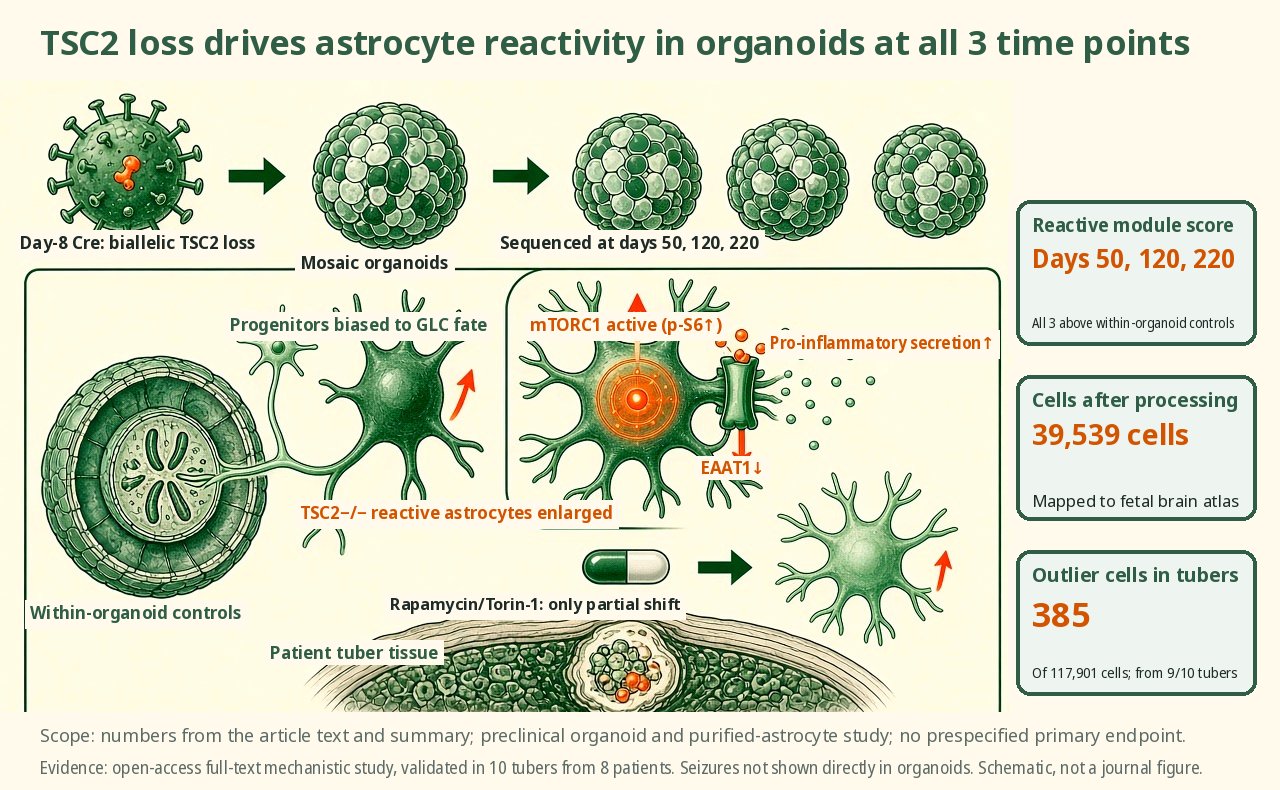
After biallelic loss of tuberous sclerosis complex 2 was induced with a recombinase lentivirus on day 8, the deleted cells in human brain organoids showed significantly higher reactive astrocyte module scores than control cells within the same organoid at days 50, 120 and 220. After processing, 39,539 cells were mapped onto a fetal brain atlas. Purified astrocytes were treated with 50 nM rapamycin or 100 nM torin. The reactive phenotype is driven cell-autonomously by mechanistic target of rapamycin complex 1.
Key data card
- Study type: Open-access full-text mechanistic study; human brain organoids, purified astrocytes and validation in patient tuber tissue
- Sample size n: 39,539 cells from WIBR3 mosaic organoids; 5 batches of BJ hiPS organoids; 10 tubers from 8 patients, with 117,901 cells in the imaging UMAP
- Controls: TSC2 c/− cells within the same organoid, isogenic TSC2+/+ organoids, TSC2 c/+ single-hit models, low p-S6 cells within tubers
- Intervention/dose: Cre lentivirus on day 8 to induce biallelic TSC2 loss; purified astrocytes treated with 50 nM rapamycin or 100 nM Torin-1
- Follow-up: scRNA-seq at 50, 120 and 220 days and at day 140; 7 days of drug treatment
- Primary endpoint: Core readout (preclinical study, no prespecified primary endpoint): reactive astrocyte module score in TSC2−/− GLCs relative to TSC2 c/− GLCs within the same organoid
- Primary endpoint result: TSC2−/− GLCs scored significantly higher than TSC2 c/− cells at 50, 120 and 220 days; the figure in the title refers to these 3 sequencing time points
- Statistics: MAST for differential expression; Wilcoxon rank-sum with Bonferroni correction for 4i intensity differences; the study is not powered as a clinical between-group comparison
- Evidence level: Full text
- Verification record: Nature open-access full-text HTML; covering the Abstract, Results, Methods, Discussion and all figure legends
- Built mosaic brain organoids
- Tracked the fate of mutant cells
- Measured reactivity proteins and secretion
- Validated in patient tuber tissue
Background and open questions
TSC is characterized by TSC1 or TSC2 mutations, cortical tubers and refractory early-onset epilepsy. Resected tuber tissue commonly shows dysmorphic neurons, gliotic astrocytes, activated microglia and giant cells, but chronic seizures themselves can also induce glial reactions, making it hard to tell whether glial changes are cause or consequence.
This paper's approach is to place biallelic TSC2 loss inside human brain organoids that never seize. The authors let mutant progenitors develop alongside TSC2 c/− cells in the same organoid, then validated the findings in patient tuber tissue, asking whether mTORC1 is sufficient to push cells into a reactive astrocyte state.
Study design
The core model is WIBR3 TSC2 c/−;LSL-TdTom human embryonic stem cell organoids. On day 8, Cre lentivirus deleted the remaining TSC2 allele in a subset of neural progenitors; tdTomato-positive cells are TSC2−/−, while tdTomato-negative TSC2 c/− cells serve as controls within the same organoid; single-cell RNA sequencing was performed at 50, 120 and 220 days.
The study is not a clinical between-group power design, nor a randomized animal experiment; the Methods state that sample sizes followed pilot experiments and the literature, that experimenters were not blinded to genotype, and that image analysis was automated. Validation included TSC2 c/+ single-hit models, 5 batches of BJ hiPS cells sequenced at day 140, full TSC2 or TSC1 knockout organoids, purified astrocytes and 4i staining of patient tubers.
Key results
The main readout holds
Across organoids and patient tubers, TSC2 loss biased progenitors toward forming enlarged, pro-inflammatory reactive astrocytes. The primary readout is the GLC reactive astrocyte module score; TSC2−/− GLCs scored significantly higher than TSC2 c/− cells in the same organoid at 50, 120 and 220 days. The module is computed from genes upregulated in reactive astrocytes, and on this basis the paper attributes the reactive signature to a cell-autonomous consequence of TSC2 loss.
A fate bias emerges
In the WIBR3 model, after Cre on day 8, 39,539 processed cells were mapped onto a fetal brain atlas. TSC2−/− cells were biased toward GLC fates relative to TSC2 c/− cells, a bias already visible at day 50 that strengthened over time. GLCs here encompass glial intermediate progenitors, oligodendrocyte progenitors and differentiated astrocytes.
Controls and replication
Controls within the same organoid rule out differences in culture conditions. TSC2 c/+ single-hit organoids showed only mild changes in proportions and expression at 50 and 120 days, with weaker reactivity than TSC2−/−; BJ male hiPS organoids, across 5 batches with Cre on day 8 and sequencing at day 140, showed the same GLC bias and reactive astrocytes.
Functional protein changes
At the protein level, TSC2−/− organoids showed increased GFAP, S100β, clusterin, CRYAB, Cx43 and Kir4.1 and strongly reduced EAAT1; this EAAT1 decrease was confirmed again in HepaCAM-purified astrocytes. Measuring 105 factors in media from 2 hiPS lines at day 280, TSC2−/− organoids secreted more GDF15, CHI3L1, CCL2, SERPINE1, HGF, CSF1, MIF and IGFBP2, with mRNA also upregulated for 6 of them.
Correspondence with lesion tissue
Purified cells at days 315–355 treated for 7 days with 50 nM rapamycin or 100 nM Torin-1 shifted only to an intermediate state, with p-S6, clusterin, APOE and p62 falling but not returning to wild-type levels. A 4i imaging UMAP of 10 tubers from 8 patients identified, among 117,901 cells, 385 outlier cells with the highest p-S6 from 9/10 tubers, with higher reactivity proteins and lower neuronal markers.
Mechanistic interpretation
Demonstrated in the paper: Within the same organoid, TSC2−/− and TSC2 c/− cells share the culture environment; cells with biallelic TSC2 loss showed a stronger GLC bias and elevated reactivity module scores. The TSC2 c/+ single-hit model showed only mild changes with weaker reactivity, supporting cell autonomy and supporting the idea that forming tuber-like cells requires biallelic loss.
The authors also connect the transcriptional state to protein, secretion and lesion tissue: EAAT1 falls, disease-associated cytokine secretion rises, and 4i shows that high p-S6 tuber cells are enriched for S100β, vimentin, APOE and clusterin and depleted of HuC/D and MAP2.
Author hypotheses: The authors propose that although these high-mTORC1 astrocytes are a very small fraction, they may still affect surrounding neuronal networks through insufficient glutamate clearance and inflammatory secretions; elevated extracellular matrix remodelling genes may also contribute to changes in tuber texture. These points are suggested by expression and secretion data; seizures were not directly demonstrated in organoids.
Limitations and uncertainties
- Extrapolation from organoids is limited: the paper states explicitly that the model lacks microglia, vascular cells and peripheral immune cells, all of which can drive astrogliosis in injury or disease; their absence supports cell autonomy but may also alter developmental trajectories.
- Patient tuber validation used resected samples from 10 tubers in 8 patients undergoing surgery for refractory epilepsy; cells with high mTORC1 activation make up less than 1% of a tuber, so their broader impact depends on the non-cell-autonomous mechanism the authors propose rather than direct functional recordings.
- Endpoints are mostly module scores, protein intensities, cell fates and secretomes, with no recording of epileptiform network activity in the organoids. mTOR inhibition was observed for only 7 days and did not fully restore wild-type states, so long-term reversal cannot be inferred.
- Methodologically, the Methods state that experimenters were not blinded to genotype; high/low p-S6 thresholds in human tubers were set manually per sample, which could shift the boundary for the small number of outlier cells.
Clinical and industry implications
If biallelic TSC2 loss first pushes progenitors toward reactive astrocytes, rather than simply producing scar-like reactions after seizures, then targets in TSC research must broaden from neurons to glial development, glutamate transport and inflammatory secretion. Organoids provide a system for tracking disease progression independently of seizures.
This remains evidence from human-derived models and resected tissue, however, and is not the same as showing in patients that suppressing astrocyte reactivity improves epilepsy. mTOR inhibition shifted cells only to an intermediate state, suggesting that lowering mTORC1 alone may not fully reset the state of lesion cells.
Authors, source and verification
Evidence level: Full text; verification record: Nature open-access full-text HTML; covering the Abstract, Results, Methods, Discussion and all figure legends
Li TL, Blair JD, Yoo T, Grant GA, Hockemeyer D, Porter BE, et al. mTORC1 drives cell-autonomous astrocyte reactivity in tuberous sclerosis. Nature. 2026. https://doi.org/10.1038/s41586-026-11054-w
Primary field: Organoids · Related: TSC2, mTORC1, Single-cell transcriptomics, 4i iterative immunostaining, Reactive astrocytes
Summary of a published paper or preprint, written from the original text; numbers are as reported by the authors. Not medical or investment advice. Corrections: contact@
Related science
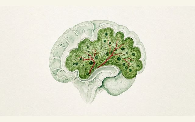
Human cortical organoids in apallial mice: grafts make up 91.9% of cortical volume
The green area in a brain cross-section is the transplanted human cortical organoid
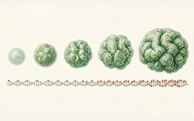
Cortical organoids at 5 years: methylation clocks track time with r=0.88–0.90
Organoids growing progressively larger, with red dots on the DNA strand below marking methylation sites
Transient confinement culture gives small intestinal organoids a 100% engraftment rate with early transplantation
Ring-shaped small intestinal organoids in a dish, with one on the right engrafted into the intestinal wall
One email, with links to every paper. Reports and custom landscapes: contact@inlightbio.com.