A GPR15 homing axis positions regulatory CD8 T cells in the gut, with a 42% reduction in sporadic UC
Human genetics, biopsies and a mouse DSS model show that GPR15 guides CD8+ TIGR homing to the colon and limits intestinal inflammation; this population was reduced by 42% in colonic biopsies from sporadic UC.
The discovery cohort comprised 7 individuals with IBD from 4 unrelated families, confirming that biallelic loss-of-function GPR15 variants cause severe colitis. A 2.5% DSS mouse model confirmed that GPR15 guides CD8+ TIGR homing to the colon and limits intestinal inflammation. This cell population was reduced by 42% in colonic biopsies from sporadic UC (HC n=8, UC n=7). GPR15-deficient mice had fewer intestinal CD8+ T cells and worse colitis after DSS. Human genetics and mouse models together support a protective role for the GPR15–TIGR axis in colonic immune homeostasis. Most main mouse experiments used n=11 per group.
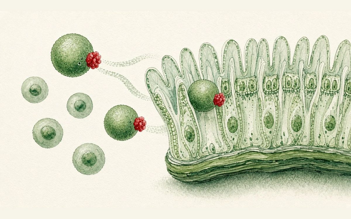
Key data card
- Study type: Human genetics, cohort association, single-cell omics and mouse mechanistic study
- Sample size n: Discovery cohort of 7 individuals with IBD from 4 unrelated families; association cohorts of 647 people (74 with IBD) and 1,692 people (109 with IBD); most main mouse experiments used n=11 per group
- Controls: Healthy controls, non-IBD controls, WT or Gpr15fl/fl mice, CD8+ splenic T cells, PBS liposomes and others
- Intervention/dose: Functional reconstitution of GPR15 variants, GPR15L migration assays, 2.5% DSS colitis, conditional Gpr15 deletion in CD8 or Foxp3 lineages, FasL/TWEAK blockade
- Follow-up: 6 days of DSS in drinking water followed by 6–7 days of plain water; some pathology readouts at day 9 or day 10; adoptive transfer for 2 weeks
- Primary endpoint: Whether GPR15 deficiency causes a homing defect in intestinal mucosal CD8+ TIGR/CD8+ T cells and weakens control of DSS colitis and macrophage inflammation
- Primary endpoint result: CD8+ TIGR as a fraction of CD8+ T cells were reduced 42% in UC colonic biopsies (HC n=8, UC n=7, two-sided Mann–Whitney U-test); after 2.5% DSS, Gpr15−/− mice lost more weight and had worse inflammation and colon shortening than WT, with Gpr15+/− intermediate
- Statistics: Mouse sample sizes were not set by statistical prespecification; figure legends report two-way ANOVA, one-way ANOVA, Kruskal–Wallis, Mann–Whitney U-test, t-test, Wald tests and FDR correction
- Safety: Mice were euthanized at the prespecified humane endpoint of >25% loss of initial body weight; patient treatment responses are discussion material, not an interventional safety trial
- Evidence level: Full text
- Verification record: The full text came from Europe PMC fullTextXML PMC13518229; materials included the Abstract, Results, Methods, figure captions, Discussion and Limitations
- Deleterious GPR15 variants weaken GPR15L chemotaxis
- CD8+ TIGR struggle to home to the colon
- Macrophages accumulate and activate after DSS
- FasL/TWEAK mediate contact-dependent killing
Background and open questions
Inflammatory bowel disease can be driven jointly by genetics and environment, and early-onset cases are especially suited to the search for monogenic clues. More than 90 genes have been linked to early-onset IBD, but the specific role of GPR15 in human intestinal immune homeostasis has remained unclear; it has been implicated in homing of mouse CD4+ Tregs or effector T cells and described as a receptor responding to GPR15L.
This paper's approach is to connect early-onset IBD families, the function of patient variants, and mouse homing and colitis models. The key question is not to show in general terms that GPR15 is "anti-inflammatory" but to establish which class of T cell it brings to the colonic mucosa and how those cells restrain myeloid inflammation after injury.
Study design
The human discovery work began with 7 individuals with IBD from 4 unrelated families, using WES to look for rare protein-altering variants; most patients had childhood onset. The study then tested GPR15 variant associations in Turkish/Qatari family cohorts, an Iranian cohort and resources such as AoU, and used 293T, HeLa, CHO-K1, Jurkat and patient T cells to measure surface expression, Ca2+ flux and GPR15L migration.
The mechanistic work used patient biopsies, public and in-house scRNA-seq, Gpr15−/− and conditional knockout mice, adoptive transfer and intestinal organoid co-culture to validate homing. Per the Methods, the DSS model used 2.5% DSS in drinking water for 6 days followed by 6–7 days of plain water; sample sizes were not prespecified by statistical calculation, and key between-group differences were tested as stated in the figure legends.
Key results
Variants associated with IBD
The Turkish/Qatari family cohort included 647 people, 74 with IBD; the Iranian cohort included 1,692 people, 109 with IBD. Compared with non-IBD controls, the authors identified 18 rare monoallelic GPR15 variants with MAF<0.001 that were significantly associated with IBD. The discovery cohort also showed clinical manifestations in 3/4 biallelic carriers and 4/10 monoallelic carriers, indicating incomplete penetrance.
Variants impair homing
Functional experiments link the variants to homing defects. WT and F159I had the highest surface expression in CHO-K1, while D306N, Y132S and Y132S/F159I were reduced and Q281X and Y215X were absent; GPR15L-induced Ca2+ influx generally tracked the decline in surface expression, with F159I an exception. Migration of patient T cells toward GPR15L was reduced in WT/M and further reduced in M/M carriers; in Pt 1.1, WT GPR15 restored surface expression and migration, while D306N and Q281X rescued minimally or not at all.
The deficit points to CD8
The core tissue-level readout is not a CD4+ Treg deficit. On the contrary, duodenal and colonic biopsies from 3 patients had a higher ratio of FOXP3+ Tregs to CD4+ T cells; colonic biopsies showed marked reductions in CD8+ T cells in both the epithelium and the lamina propria. The GPR15–GPR15L axis therefore guides a class of colonic CD8+ T cells in humans rather than the CD4+ Treg compartment.
DSS localizes the protective cells
After 2.5% DSS, Gpr15−/− mice lost weight faster and had worse inflammation and more pronounced colon shortening, with Gpr15+/− showing intermediate changes. Conditional knockouts localized this further to CD8+ T cells: Gpr15fl/fl Cd8cre worsened DSS colitis while Gpr15fl/fl Foxp3cre did not. The main mouse groups in Fig. 4 used n=11 per group and were tested by ANOVA, Mann–Whitney or similar.
Macrophages are killed
The immune cell readouts point to macrophages rather than dendritic cell expansion. At day 3 of DSS, F4/80+ macrophages increased in Gpr15−/− colon while CD103+ dendritic cells did not; by day 5 macrophages stained strongly for NOS2 and CD206. In vitro co-culture showed that blocking FasL or TWEAK reduced killing, with combined blockade comparable to anti-FasL alone, indicating that FasL predominates. CD8+ TIGR were reduced 42% in sporadic UC and also significantly reduced in the terminal ileum in Crohn's disease.
Mechanistic interpretation
Demonstrated in the paper: The paper directly shows that several deleterious GPR15 variants reduce receptor surface expression, Ca2+ signalling and GPR15L chemotaxis, with F159I an exception for surface expression and migration; patient T cells agreed with the Jurkat and CHO-K1 results, and reconstitution with WT GPR15 in Pt 1.1 rescued migration while D306N and Q281X did not. Patient biopsies and Gpr15−/− mice both showed loss of colonic CD8+ T cells, with no corresponding deficit in CD4+ FOXP3+ Tregs.
The experiments support each step, but the paper presents the overall claim that "CD8+ TIGR kill inflammatory macrophages through FasL/TWEAK to prevent colitis" as a suggestive model: after 2.5% DSS, colitis worsened in Gpr15−/− mice and CD8-specific but not Foxp3-specific deletion reproduced the phenotype; F4/80+ macrophages accumulated and stained strongly for NOS2/CD206. Activated CD8+ TIGR directly killed LPS-activated macrophages in vitro, and blocking FasL or TWEAK reduced killing, so the death receptor mechanism rests mainly on in vitro blockade.
Author hypotheses: The authors suggest that a reduction in CD8+ TIGR may weaken local restraint on macrophage-driven inflammation in sporadic UC and Crohn's disease, and that CD8+ TIGR TCRs may recognize self, dietary or microbially derived antigens, though those antigens were not identified here. They also propose that enhancing the GPR15 pathway or controlling pathogenic macrophage activity could become a therapeutic direction in IBD.
Limitations and uncertainties
- First, genetic extrapolation is limited by incomplete penetrance. The population frequency of Y132S constrains a monogenic explanation: MAF=0.0012, with 1,916 heterozygotes and 20 homozygotes in gnomAD v.4.1.1, so the authors interpret it as a hypomorphic allele rather than a fully penetrant pathogenic variant.
- Second, the discovery cohort comprised only 7 individuals with IBD from 4 unrelated families; variant carriers were clinically heterogeneous, with penetrance of 3/4 for biallelic and 4/10 for monoallelic carriers. The sporadic UC analysis used HC n=8 and UC n=7 and the Crohn's analysis HC n=8 and CD n=20, which can indicate association but cannot substitute for prospective risk estimates.
- Third, mouse sample sizes were not prespecified by statistical calculation and investigators were not blinded to genotype groups; the DSS model used 2.5% DSS to cause acute epithelial injury with follow-up to day 9 or day 10, which cannot cover chronic relapse or long-term repair. Fourth, the paper does not identify the antigen specificity of CD8+ TIGR, nor does it provide dosing or a safety window for GPR15-targeted therapy.
Clinical and industry implications
If later cohorts confirm the relationship between GPR15 variants, CD8+ TIGR deficits and clinical subtypes, this work could move part of IBD from "generalized mucosal inflammation" toward mechanistic stratification by "impaired homing and uncontrolled macrophages". As a membrane receptor, GPR15 could also become a diagnostic or interventional node.
These conclusions should stay within the paper's evidence, however: patient material supports the genetic association and the tissue deficit, and mouse and in vitro experiments support the causal mechanism; the 42% and the significant reductions in sporadic UC and Crohn's disease remain associations in single-cell datasets and do not show that enhancing CD8+ TIGR has been validated to benefit patients.
Authors, source and verification
Evidence level: Full text; verification record: The full text came from Europe PMC fullTextXML PMC13518229; materials included the Abstract, Results, Methods, figure captions, Discussion and Limitations
Cui J, Chen Z, Cheng YH, Nie J, Zhu MH, Xiang C, et al. GPR15-guided CD8+ T regulatory cells control intestinal inflammation. Nature. 2026. https://doi.org/10.1038/s41586-026-10749-4
Primary field: Autoimmunity & transplant · Related: GPR15-GPR15L, CD8+ regulatory T cells, Inflammatory bowel disease, DSS colitis, FasL-TWEAK
Summary of a published paper or preprint, written from the original text; numbers are as reported by the authors. Not medical or investment advice. Corrections: contact@
Related science
Anti-CD19 CAR-T in refractory MG: 3 patients in treatment-free remission for at least 19 months
The neuromuscular junction at left, CAR-T cells clearing red B cells at right
Two highly sensitized patients transplanted after dual-target CAR-T
Five green CAR-T cells and one red target cell, with a kidney at right
Hashimoto's thyroiditis: 135 distinct TNFRSF14 mutations in a single donor
Thyroid follicles at left, B cell clones at right, with red dots marking mutations
One email, with links to every paper. Reports and custom landscapes: contact@inlightbio.com.