A pIgR bispecific raises exposure in cynomolgus monkey BAL 5.5-fold
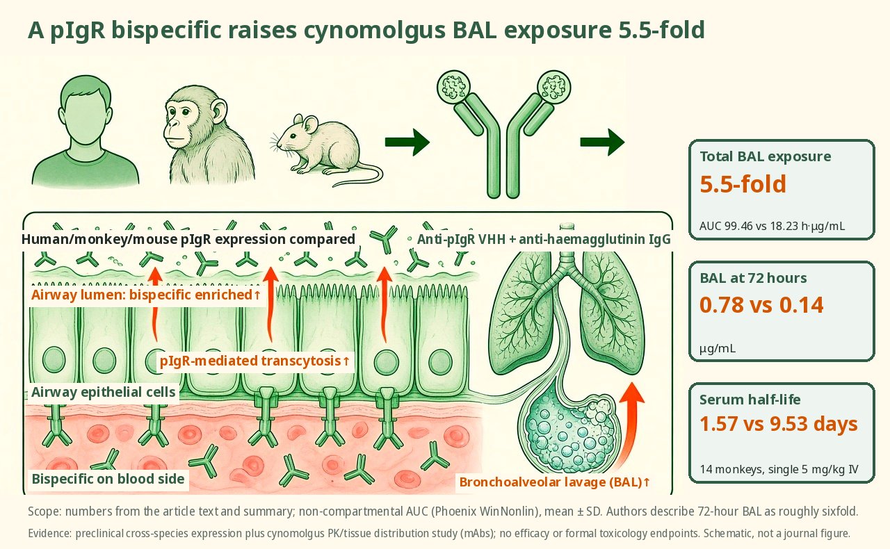
After comparing the polymeric immunoglobulin receptor (pIgR) across human, cynomolgus monkey and mouse tissues, Marks et al. fused an anti-pIgR VHH onto an anti-haemagglutinin IgG; after a single intravenous dose of 5 mg/kg in cynomolgus monkeys, the bispecific had a serum half-life of about 1.6 days and total exposure in bronchoalveolar lavage (BAL) fluid 5.5 times that of the control IgG.
After comparing the polymeric immunoglobulin receptor across human, cynomolgus monkey and mouse tissues, an anti-pIgR VHH was fused onto an anti-haemagglutinin IgG. In 14 cynomolgus monkeys given a single intravenous dose of 5 mg/kg, the bispecific had a serum half-life of 1.57 days (9.53 days for the control) and total bronchoalveolar lavage exposure 5.5 times that of the control (AUC 99.46 versus 18.23 h·µg/mL), with 72-hour concentrations of 0.78 versus 0.14 µg/mL. Mucosal pIgR transcript profiles were similar in humans and cynomolgus monkeys, while high expression in mouse liver constitutes a species difference, so primates were chosen for the pharmacokinetic study.
Key data card
- Study type: Preclinical bispecific delivery study combining cross-species expression profiling with pharmacokinetics and tissue distribution in cynomolgus monkeys (mAbs)
- Sample size n: RNA-seq in 5 female and 5 male mice; cynomolgus monkeys were sexually mature animals of Chinese or Cambodian origin (the paper notes 5 male and female animals without further breakdown); IHC on human tissue arrays of 180 and monkey arrays of 240 (n≥3 per site); pharmacokinetics in 14 cynomolgus monkeys (8 on FM1B104, 6 on the control FM1B108; serum n=4 and n=3 in Table 1)
- Controls: An IgG with the same haemagglutinin arm and no pIgR VHH (FM1B108); a control antibody that does not bind haemagglutinin
- Intervention/dose: A single intravenous dose of 5 mg/kg; FM1B104 is an anti-haemagglutinin IgG with anti-pIgR VHH2 fused to the heavy chain N terminus
- Primary endpoint: Serum and BAL pharmacokinetic parameters; tissue concentrations in lung, kidney and duodenum/jejunum/ileum
- Primary endpoint result: BAL AUC_all of 99.46 versus 18.23 h·μg/mL (5.5-fold); BAL at 72 hours of 0.78 versus 0.14 μg/mL (described by the authors as roughly sixfold); serum t½ of 1.57 versus 9.53 days
- Statistics: Non-compartmental modelling (Phoenix WinNonlin); linear up/log down trapezoidal AUC; mean ± SD
- Safety: This is a PK/tissue distribution study with no efficacy or formal toxicology endpoints
- Evidence level: Full text
- Verification record: Europe PMC full-text XML (PMC13644599): Abstract, Results, Methods, Discussion, Tables 1–2 and legends for Figs. 1–6
- Cross-species confirmation of mucosal pIgR expression
- An anti-pIgR VHH fused to the heavy chain N terminus
- Faster serum clearance with higher BAL exposure
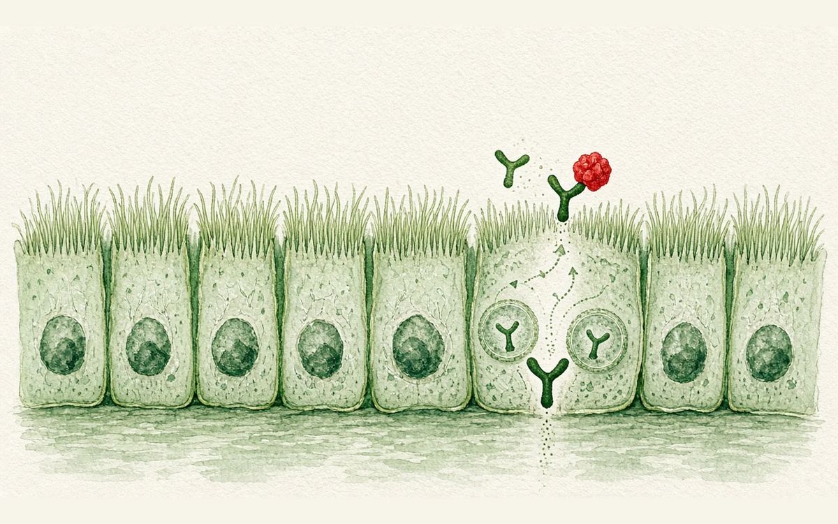
- Antibody in mucosal tissue is transported away
Background and open questions
IgG's inability to reach the mucosal lumen is a long-standing problem for antibody drugs. The paper cites that therapeutic IgG exposure in lung mucus is only about one thousandth of serum, that haemagglutinin antibodies often require doses of around 50 mg/kg, and that humans transport roughly 3–5 g of dimeric IgA per day through pIgR. pIgR sits mainly on the basolateral side of mucosal epithelium, and after binding dIgA it transcytoses to the apical side and is cleaved off, forming secretory IgA. Targeting pIgR with peptides, VHHs or dIgA has already produced directional transcytosis in cells and human microtissues, but high pIgR in rodent liver skews pharmacokinetics through hepatic clearance.
Marks and Shah (equal contributors) with Cheung and Zwolak (corresponding) narrow the question to two steps: whether expression runs in the same direction in human, cynomolgus monkey and mouse; and whether the same anti-haemagglutinin backbone, once fused to a previously characterized anti-pIgR VHH, genuinely enriches in cynomolgus lung and gastrointestinal tract.
Study design
Transcriptomes were compared by TPM. Mice were C57BL/6J at about 7–8 weeks, 5 female and 5 male, merged with public datasets; cynomolgus monkeys were sexually mature animals of Chinese or Cambodian origin (the paper notes 5 male and female animals without further breakdown). Protein was assessed by IHC with the rabbit monoclonal EPR23314-256, with cell block controls plus human 180-core and monkey 240-core tissue arrays (n≥3 donors per site). Human and monkey pIgR proteins are 89% identical.
On the antibody side, FM1B108 is a monoclonal carrying the CR9114 haemagglutinin variable regions; FM1B104 fuses anti-pIgR VHH2 to its heavy chain N terminus. A control that does not bind haemagglutinin was also included. Binding was measured by biolayer interferometry, with antibody and antigen at 10 μg/mL per the figure legends. Pharmacokinetics: 14 cynomolgus monkeys received a single intravenous dose of 5 mg/kg, 8 given FM1B104 and 6 FM1B108; serum time points included pre-dose and 0.5 to 312 hours; BAL used 10 mL of saline without correction for epithelial lining fluid dilution; lung, kidney and three small intestinal segments were taken at 48 and 312 hours. Concentrations were measured by MSD with a lower limit of quantification of 5 ng/mL. Parameters were derived by non-compartmental analysis. The BAL/serum counts of 4 and 3 in Table 1 reflect staggered sampling and scheduled necropsies, so fewer animals than enrolled contributed full curves.
Key results
High in mouse liver, consistent across primate mucosa
Most digestive tissues in mice exceeded 500 TPM, with prostate and uterus above 40 TPM and liver also above 500 TPM. In cynomolgus monkeys, colon and stomach exceeded 500 TPM, while jejunum, liver, cervix, mammary gland and kidney were below 500 TPM. In humans, salivary gland, colon, jejunum and smooth muscle exceeded 500 TPM, while kidney, lung, ileum, stomach, pancreas and mammary gland were below 500 TPM. The largest species difference is in liver: high in mouse and very low in cynomolgus monkey and human (the Discussion gives about 14 TPM for humans and about 23 TPM for monkeys). IHC showed membrane/cytoplasmic positivity in human duodenal mucosa, renal tubules and bladder epithelium, with hepatocytes essentially unstained and apical staining in bile ducts; comparable sites stained in cynomolgus monkeys but more weakly than in humans, with liver negative. The authors state explicitly that, apart from renal tubules, midbrain neurons and tonsil/lymph node leukocytes, staining patterns are not identical between human and monkey, and non-specific antibody binding in monkey is possible. In lung, humans showed positive alveolar macrophages while monkeys showed rare alveolar macrophages and apical tracheal epithelium.
Only the VHH-bearing molecule binds pIgR
Both FM1B104 and FM1B108 bind H1N1 haemagglutinin, but only the former binds human pIgR. Among controls, B23B307 binds both human and cynomolgus pIgR, while MSCB847 binds neither. Subsequent pharmacokinetics therefore compared FM1B104 with FM1B108, attributing the differences mainly to the pIgR arm.
Higher BAL exposure with a shorter serum half-life
FM1B104 had a terminal serum half-life of 1.57±0.5 days versus 9.53±0.5 days for the control (rounded to 1.5 and 9.5 days in the Results narrative). BAL Cmax was 0.78±0.4 μg/mL for FM1B104 versus 0.14±0.1 μg/mL for the control. The Results state that both antibodies peaked in BAL at 72 hours, while Table 1 gives Tmax of 60±24 and 104±55 hours. BAL AUC_all was 99.46±35.2 versus 18.23±6 h·μg/mL, a 5.5-fold difference. Serum/BAL ratios at 72 hours were 28 versus 396, which the authors take as roughly a sixfold window at peak. The Introduction states that the high BAL exposure persists for about a week, consistent with the mucosal half-life of dIgA. Clearance was 0.9±0.3 versus 0.2±0.03 mL/h/kg (described in the Discussion as roughly 4.5-fold) and volume of distribution 54.8±37 versus 72.8±13 mL/kg, so the difference lies mainly in clearance rather than broad distribution. The paper cites healthy adult values of 11 mg/mL IgG in serum and 0.0092 mg/mL in BAL (a ratio of 1195) and 1.9 and 0.013 mg/mL for sIgA (a ratio of 146), to argue that the bispecific's mucosal ratio is better than steady-state IgG/IgA. BAL was not corrected for dilution, so concentrations should be compared only in relative terms.
The bispecific falls faster in tissues
Lung tissue concentrations at 48 hours were similar (12.16±5.69 versus 10.14±2.12 ng/mg), while at 312 hours the bispecific fell to 0.13±0.08 ng/mg against 4.08±1.67 ng/mg for the control, described in the Results as nearly 30-fold lower. Duodenum, jejunum and ileum were similar between the two at 48 hours, with the bispecific below the limit of quantification in all three at 312 hours while the control remained measurable. Kidney already showed lower bispecific levels at 48 hours (0.95±0.28 versus 3.23±1.61 ng/mg, about 3.4-fold) and was likewise below the limit of quantification at 312 hours. The authors link this faster renal clearance to the uniform pIgR staining of primate renal tubules.
Mechanistic interpretation
Demonstrated in the paper: pIgR transcript and mucosal epithelial protein patterns are similar in human and cynomolgus monkey, while high expression in mouse liver constitutes a species trap, giving a tissue-level rationale for not using mice in transcytosis pharmacokinetics. Adding a pIgR VHH to the same haemagglutinin arm shortened serum half-life, raised BAL AUC and drove pIgR-positive tissues below the limit of quantification at late time points, consistent in direction with earlier transcytosis data in cells and human microtissues. Clearance rose while volume of distribution was similar, supporting receptor-related disposition rather than a larger systemic volume.
Author hypotheses: Absolute BAL concentrations were not corrected for epithelial lining fluid and cannot be taken directly as true mucosal fluid values; mechanistically it is also not yet possible to quantitatively separate transcytosis, target-mediated clearance and other disposition. Anti-haemagglutinin was chosen to minimize target-mediated clearance through a second target in healthy animals. The Discussion proposes that rapid clearance from blood might also be used to remove pathogenic IgG, an extrapolation rather than a readout from this experiment. Declines in kidney and gastrointestinal tissue are interpreted as antibody having been transported to the luminal surface, but the study did not directly measure intestinal lumen or cyst fluid.
Limitations and uncertainties
- IHC was weaker overall in monkeys than in humans, and the authors acknowledge that staining patterns are not identical outside a few sites, with possible species non-specificity; strong bladder IHC despite low transcript levels is attributed to a small fraction of expressing cells and was not separately validated.
- Pharmacokinetic n values are smaller than enrolment n, and BAL was not corrected for dilution; the study was not designed to distinguish transcytosis from other clearance routes, and includes no infection or cyst efficacy endpoints.
- Lung pIgR RNA is lower in cynomolgus monkeys than in humans, so the authors suggest that enrichment in monkeys may underestimate humans, while the reverse holds for colon. Mice were explicitly excluded because of their distinctive liver and oesophageal expression. Earlier data on anti-EGFR dIgA in polycystic kidney models come from the literature and cannot be taken as validation of this bispecific in cyst fluid.
Clinical and industry implications
If human pharmacokinetics point the same way, pIgR bispecifics could deliver IgG to the lung mucosa at lower systemic doses and shorten the onset time of antibodies against acute respiratory infections; they also offer an IgG-based alternative to dIgA manufacturing for indications that need luminal access while avoiding systemic growth factor receptor engagement, such as polycystic kidney disease. The cost is a short serum half-life and brief residence in extrahepatic mucosal tissue, so dosing cadence for chronic administration would differ from ordinary IgG. The next steps require disease models and formal safety assessment rather than writing relative BAL fold changes directly into clinically effective concentrations.
Authors, source and verification
Equal contributors: Kelsie Marks and Nirav Shah; last authors: Wan Cheung Cheung and Adam Zwolak (corresponding). Evidence level: Full text; verification record: Europe PMC full-text XML (PMC13644599): Abstract, Results, Methods, Discussion, Tables 1–2 and legends for Figs. 1–6. The licence is CC BY-NC, so the text is a paraphrase without sentence-by-sentence reproduction or use of original figures.
Marks K, Shah N, Pham L, Chen J, White I, Burke K, et al. Bispecific antibodies that engage pIgR enrich in mucosa in cynomolgus monkeys. mAbs. 2026 Oct 3;18(1):2743413. doi: https://doi.org/10.1080/19420862.2026.2743413
Primary field: Antibody engineering · Related: Disease models
Summary of a published paper or preprint, written from the original text; numbers are as reported by the authors. Not medical or investment advice. Corrections: contact@
Related science
A 156-member bispecific screen selects a low-affinity SIRPα×CD38
A macrophage phagocytosing the tumour cell at right via a red bispecific
A trispecific IL-2R antibody reaches an EC50 of 0.015 nM with Treg bias
A green trispecific antibody binding a dark green regulatory T cell, with the rest quiescent
A 160-member bispecific library: surface properties predictable from parents, ρ up to 0.95
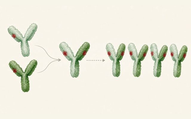
Two parental antibodies combining into a bispecific, with red dots marking surface properties
One email, with links to every paper. Reports and custom landscapes: contact@inlightbio.com.