Charge-switchable lipid nanoparticles did not raise IL-6, IL-1β or MIP-2 in LPS-pretreated mice
In LPS-pretreated mice, E20 switchable lipid nanoparticles (SNPs) did not raise IL-6, IL-1β or MIP-2, while LNPs made with MC3-DLin, cKK-E12 or SM-102 raised them sharply.
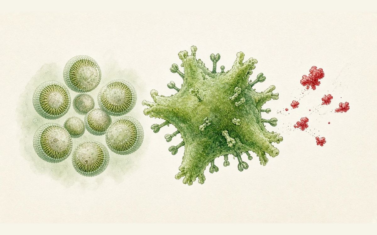
When mice pretreated with 1 mg/kg LPS received 1 mg/kg mRNA 4 hours later, the E20 charge-switchable LNP (SNP) did not raise IL-6, IL-1β or MIP-2, while conventional LNPs made with MC3-DLin, cKK-E12 or SM-102 raised them sharply, making the SNP the only LNP that did not worsen existing inflammation. With pDNA at 1 mg/kg, all mice given MC3-DLin LNPs died within 24 hours, whereas all mice in the E20-SNP group survived the 7-day observation period with sustained luciferase expression. In human PBMC experiments (4 healthy donors), E20-SNP transfected efficiently and induced none of 14 cytokines. E20-SNP activated neither complement, TLR4, galectin-8 nor the PAF pathway, and its zeta potential was more negative than that of MC3-DLin.
Key data card
- Study type: Preclinical study (in vitro cells, human PBMCs, mouse models)
- Sample size n: HEK cell screening n=3; mouse luciferase delivery n=2; hEPO, LPS pretreatment, pDNA and lung injury each n=3; human PBMCs from 4 healthy donors
- Controls: MC3-DLin, SM-102, ALC-0315, 4A3-SC8 and cKK-E12 LNPs; PBS, untreated, LPS alone and an E20-SNP/mLuc scrambled mRNA group
- Intervention/dose: E20-SNP (an LNP made from an S-lipid); 1 mg/kg mRNA given 4 h after 1 mg/kg LPS; pDNA at 1 mg/kg; IL-22 mRNA at 2.5 mg/kg intratracheally
- Follow-up: Cytokines at 4 and 24 h; pDNA over 7 days; lung injury 24 h after treatment
- Primary endpoint: Central readout: serum IL-6, IL-1β and MIP-2 in LPS-pretreated mice (E20-SNP versus several conventional LNPs); co-primary readouts are survival with pDNA and LPS lung injury measures
- Primary endpoint result: E20-SNP did not raise IL-6, IL-1β or MIP-2 and was the only LNP that did not worsen existing inflammation; MC3-DLin, cKK-E12 and SM-102 LNPs raised them sharply (Fig. 3f, ANOVA + Dunnett; no specific values given in the text). With pDNA at 1 mg/kg, all MC3-DLin animals died within 24 h while the E20-SNP group survived 7 days
- Statistics: One-way ANOVA with Dunnett/Tukey/Holm-Šídák, log-rank test for survival, data as mean ± s.e.m.
- Safety: Mice given E20-SNP looked comparable to untreated controls; conventional LNP groups showed hunching, reduced activity and squinting; all mice in the MC3-DLin pDNA group died
- Evidence level: Full text
- Verification record: Read the abstract, Main, all Results subsections, legends for Figs. 1–6 and Extended Data Figs. 1–3, Methods and Conclusion of the open-access Nature Nanotechnology full text
- Screening of 144 S-lipids, with the top 18 validated
- SNPs are negatively charged at pH 7.4 and deliver mRNA/pDNA
- E20-SNP activated neither complement, TLR4, galectin-8 nor PAF
- LPS-pretreated mice: IL-6 and other cytokines did not rise
- pDNA: all animals in the LNP group died within 24 h while the SNP group survived 7 days
Background and open questions
LNP–mRNA complexes offer transfection efficiency and tissue tropism, but clinical translation beyond vaccines has been slowed by toxicity. That toxicity arises from several inflammatory pathways including complement, TLR4 and galectin signalling, and LNPs also trigger release of platelet-activating factor (PAF), which is more pronounced in patients with existing inflammation.
The difficulty is that the positive charge of ionizable lipids and their ability to disrupt endosomes are both the main source of toxicity and necessary for binding mRNA, forming a protein corona and triggering endosomal release, so conventional LNPs trade toxicity against delivery efficiency. This study (Nature Nanotechnology) designs S-lipids, ionizable lipids whose head groups contain a carboxyl and a tertiary amine and that are negatively charged at pH 7.4, and calls the resulting particles SNPs, testing whether they can avoid these pathways while preserving delivery.
Study design
This is a preclinical study comprising in vitro screening and mouse and human PBMC experiments. First, 144 S-lipids were synthesized and tested for luciferase mRNA transfection in HEK cells (500 ng/ml, 24 h, n=3), and the top 18 were then purified and validated in HEK cells and primary mouse fibroblasts. In vivo, BALB/c mice received intravenous injections: luciferase mRNA at 0.25 mg/kg (n=2) and hEPO mRNA at 0.1 mg/kg (n=3).
The central comparison is SNPs made with the representative molecule E20 against several conventional LNPs (MC3-DLin, SM-102, ALC-0315, 4A3-SC8 and cKK-E12). In the LPS pretreatment model, LPS at 1 mg/kg was given first, followed 4 h later by 1 mg/kg mRNA, with blood drawn at 4 and 24 h for cytokines (n=3). The pDNA experiments used 3 mice per group at 1 mg/kg; the lung injury model used intratracheal LPS at 3 mg/kg followed 3 h later by IL-22 mRNA at 2.5 mg/kg. Sample sizes were 2–3 animals per group or 4 donors, with no statement of a power design for between-group comparison; test methods are given in the figure legends.
Key results
Screening and physicochemical properties
Screening of 144 S-lipids showed that 6–8 carbons are needed in the linker between head group amine and carboxyl, together with branched hydrophobic tails and internal spacers; molecules with these modifications were up to 9,500-fold more potent than those with linear tails. Among the top 18 purified molecules, SNPs made from 11 (61.1%) transfected fibroblasts as efficiently as cKK-E12 or SM-102 LNPs; their pKa values were 4.2–5.8 and their zeta potentials more negative than those of MC3-DLin LNPs.
mRNA delivery in vivo
In the in vivo evaluation of the top 18 SNPs (luciferase mRNA at 0.25 mg/kg, n=2), 5 matched or exceeded the delivery efficiency of MC3-DLin LNPs and 10 had more than 80% of their signal in the liver. With 0.1 mg/kg hEPO mRNA given intravenously, E20-SNP produced blood hEPO of 600 mU/ml, 100 times the therapeutic threshold and comparable to MC3-DLin LNPs.
Inflammation in LPS-pretreated mice
In the LPS pretreatment model (n=3), MC3-DLin, cKK-E12 and SM-102 LNPs raised serum IL-6, IL-1β and MIP-2 sharply, while E20-SNP did not raise these cytokines and was the only LNP that did not worsen existing inflammation. Mice given E20-SNP looked comparable to untreated controls, while LNP groups showed hunching, reduced activity and squinting. This readout is shown in Fig. 3f with one-way ANOVA and Dunnett tests.
pDNA toxicity and human PBMCs
After 1 mg/kg of nanoplasmid pDNA, all mice given MC3-DLin LNPs died within 24 h; the E20-SNP–pDNA group showed no distress, survived the entire 7-day observation period and continued to express luciferase (3 mice per group, log-rank test). In human PBMC experiments (4 healthy donors, 2,000 ng/ml), E20-SNP transfected efficiently and induced none of 14 cytokines, while MC3-DLin, 4A3-SC8, ALC-0315, SM-102 and cKK-E12 LNPs induced multiple inflammatory cytokines.
Treatment of LPS acute lung injury
In the lung injury model, E20-SNP fully reversed the lung permeability phenotype, with lung protein levels comparable to healthy mice and cellular infiltration comparable to untreated controls; the MC3-DLin, SM-102, ALC-0315 and 4A3-SC8 LNP groups still showed protein leakage and increased cellular infiltration (n=3 per group, Tukey test). IL-22 mRNA delivery efficiency with E20-SNP matched conventional LNPs, with an additional E20-SNP/mLuc scrambled mRNA control group.
Mechanistic interpretation
Demonstrated in the paper: C3a did not increase after human serum was incubated with E20-SNP (2,000 ng/ml, 37 °C for 1 h), with zymosan as a positive control; E20-SNP did not activate TLR4 in TLR4 reporter cells, with LPS as a positive control, while several conventional LNPs activated both. In galectin-8 reporter cells (1,000 ng/ml), E20-SNP caused only minimal galectin-8 recruitment to endosomes; it did not trigger PAF release in human PBMCs, while conventional LNPs activated both pathways.
The low toxicity is not due to insufficient transfection: luciferase expression from E20-SNP in galectin-8 reporter cells was comparable to conventional LNPs, and the luciferase signal persisted over 7 days in the pDNA mice. In haemolysis assays, E20-SNP was not membrane-disruptive at pH 5.5 while all conventional LNPs were; Cy5-mRNA tracing showed E20-SNP accumulating in Rab11+ endosomes.
Author hypotheses: The authors speculate that E20-SNP enters the cytosol through Rab11+ endosomes without activating galectin-8 or PAF, and attribute the avoidance of complement and TLR4 to a head group that is negatively charged or neutral at pH 7.4; the advantage in lung injury most likely stems from low immune stimulation rather than a difference in delivery. These attributions were not directly tested in the experiments described, for example by altering head group charge.
Limitations and uncertainties
- The authors did not study intracellular metabolism and accumulation of S-lipids, and SNPs may become toxic through accumulation after chronic dosing; future molecules may need ester bonds in the hydrophobic tails to speed clearance.
- Sample sizes are small: in vivo luciferase delivery used n=2 per group, pDNA and most mouse experiments 3 animals per group, and PBMCs 4 donors; in the pDNA experiment all 3 MC3-DLin animals died, so no blood could be drawn and serum cytokine controls for that group are missing. The text gives no specific P values for the individual comparisons.
- Follow-up and scope are limited: pDNA was observed for only 7 days and lung injury analysed only 24 h after treatment; the immunotoxicity comparisons centre on the single molecule E20, with cytokine data for other S-lipids not presented in the text; and the evidence comes from mice and in vitro human PBMCs, with no human data.
Clinical and industry implications
If these results are reproduced in more models, switchable ionizable lipids whose head groups are negatively charged at pH 7.4 may offer a route to lower LNP inflammatory toxicity for nucleic acid therapies that require high doses, repeat dosing or use in patients with existing inflammation, with the paper's pDNA delivery and lung inflammation settings as two examples.
Current evidence is limited to mice and human PBMCs, with comparator lipids including MC3-DLin, SM-102 and ALC-0315. Whether this translates to human use depends on intracellular metabolism and accumulation, chronic dosing safety, and re-examination of each inflammatory pathway in larger samples.
Authors, source and verification
Evidence level: Full text; verification record: Read the abstract, Main, all Results subsections, legends for Figs. 1–6 and Extended Data Figs. 1–3, Methods and Conclusion of the open-access Nature Nanotechnology full text
Liang D, Qi Y, Han H, Ahmadian N, Gao K, Sapasap K, et al. Charge-switching ionizable lipids lower the toxicity of lipid nanoparticles. Nat Nanotechnol. 2026. https://doi.org/10.1038/s41565-026-02262-6
Primary field: Nucleic acid & gene therapy · Related: Switchable ionizable lipids, S-lipids, LPS-pretreated mice, pDNA toxicity, galectin-8, Acute lung injury
Summary of a published paper or preprint, written from the original text; numbers are as reported by the authors. Not medical or investment advice. Corrections: contact@
Related science
A single inhaled dose of ARO-RAGE lowered BALF sRAGE by 90.2%
Alveoli and droplets at left, with the siRNA cleaving messenger RNA at right
RAG-17 phase 1 in 6 patients: cerebrospinal fluid SOD1 down 69% and 56%
A red strand inside a motor neuron cell body representing the silenced messenger RNA
A single dose of PE-LNP achieved 49% precise editing across the mouse liver
A lipid particle carrying red editing components into a hepatocyte
One email, with links to every paper. Reports and custom landscapes: contact@inlightbio.com.